Yigong Shi’s Group Elucidated the cryo-EM Structures of the Catalytically Activated Yeast Spliceosome on Cell
On March 15th 2019, Professor Yigong Shi’s group in the School of Life Sciences, Tsinghua University, published an article entitled “Structures of the Catalytically Activated Yeast Spliceosome Reveal the Mechanism of Branching” in the prestigious international magazine Cell. In this paper, Shi and colleagues assembled the catalytically activated spliceosomes (B* complex) on two different pre-mRNAs and determined the cryo-EM structures of four distinct B* complexes from Saccharomyces cerevisiae.at the overall resolutions of 2.9-3.8 A. Comparison of these structures reveals mechanistic insights into the branching reaction.
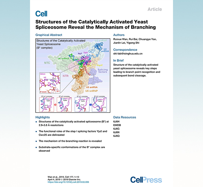
In 1977, scientists observed that the adenovirus mRNA could not form a continuous double-stranded RNA-DNA hybrid duplex with its corresponding DNA transcription template. Instead, single-stranded DNA bulges were extended at different positions in the hybridized double-stranded RNA-DNA duplex. This discovery suggested that the transfer of genetic information from DNA to mRNA involved not only transcription but also pre-mRNA splicing, which is to further remove the non-coding regions (introns) and joined the coding sequences (exons). RNA splicing is ubiquitous in eukaryotes. The average numbers of introns per gene increase from simple single-cell eukaryotes, such as yeast, to higher organisms such as worms and flies, and all the way up to humans. Some pre-mRNAs can be spliced in more than one way. Therefore, mRNAs containing different combinations of exons can be generated from a given pre-mRNA.
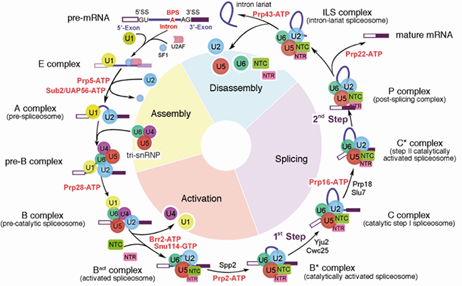
Figure 1. The precursor messenger RNA (pre-mRNA) splicing cycle.
(Yan, C., Wan, R., & Shi, Y. (2019). Cold Spring Harbor perspectives in biology, 11(1), a032409.)
Pre-messenger RNA (pre-mRNA) splicing is a crucial and the most complicated step in the central dogma in eukaryotic cells. This process is executed by a dynamic mega-complex known as the spliceosome. From the discovery of RNA splicing in 1977 to the beginning of this century, scientists have recapitulated assembly and disassembly of the spliceosome by immunoprecipitation, gene knockout, cross-linked mass spectrometry, and in vitro splicing reaction and so on. The essence of RNA splicing is the removal of intron and ligation of exons through two successive transesterification reactions in which phosphodiester linkages within the pre-mRNA are broken and a new one is formed. To catalyze splicing of the pre-mRNA, spliceosome has to undergo extremely precise stepwise assembly by forming a series of spliceosomal complexes. Based on the assembly and catalytic states and the biochemical composition of the spliceosome, these spliceosomal complexes have been defined as the A, pre-B, B, Bact , B*, C, C*, P, and ILS complexes. (Figure 1)
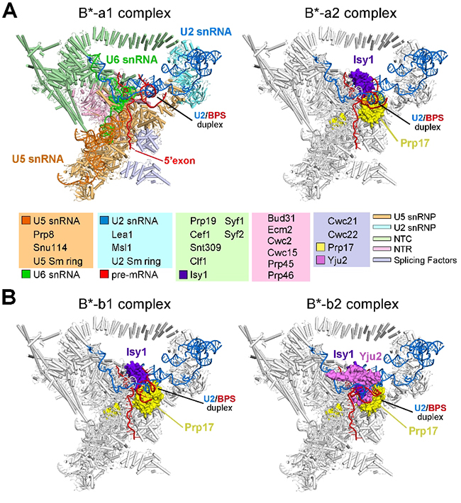
Figure 2. Cryo-EM structures of the catalytically activated spliceosomes (B* complexes) from Saccharomyces cerevisiae.
The catalytically activated spliceosome (B* complex) is a highly transient complex that is difficult to be captured under normal physical conditions. In their recent Cell paper, Shi and colleagues reconstituted the B* complex using the in vitro assembly approach, in which the assembled Bact complex was remodeled into the B* complex upon incubation with recombinant Prp2 and Spp2 in the presence of ATP. Then they acquired the good B* complexes by applying affinity purification strategy and determined the three-dimensional structures by single particle cryo-EM, and built the atomic model (Figure 2). Structure of the S. cerevisiae B* complex contains 35 discrete proteins, three snRNA molecules, and the pre-mRNA. Structural elucidation of the B* complex fills an important void in the mechanistic understanding of pre-mRNA splicing by the spliceosome. The duplex between U2 snRNA and the branch point sequence (BPS) is discretely away from the 5’-splice site (5’SS) in the three B* complexes that are devoid of the step I splicing factors Yju2 and Cwc25. Recruitment of Yju2 into the active site brings the U2/BPS duplex into the vicinity of 5’SS, with the BPS nucleophile positioned 4 A away from the catalytic metal M2. This analysis reveals the functional mechanism of Yju2 and Cwc25 in branching. These structures on different pre-mRNAs reveal substrate-specific conformations of the spliceosome in a major functional state (Figure 3).
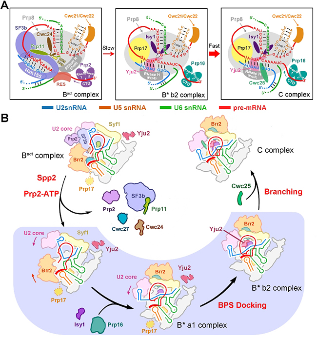
Figure 3. Mechanism of branching catalyzed by the spliceosome in S. cerevisiae.
The Shi Group has been focusing on the structural and mechanistic elucidation of pre-mRNA splicing in the past decades. After the success of crystal structural elucidation of subcomplexes within spliceosome, they took a direct assault on the intact spliceosome and made the breakthrough in the summer of 2015. Since then, the Shi lab has reported nine high resolution structures of the crucial spliceosomal complexes, including the S.pombe intron lariat spliceosome ILS complex at 3.6 A, the pre-assembled U4/U6.U5 tri-snRNP at 3.8 A, the precursor pre-catalytic spliceosome pre-B complex at 3.3-4.6 A, the pre-catalytic spliceosome B complex at 3.9 A, the activated spliceosome Bact complex at 3.5 A, the catalytic step I spliceosome C complex at 3.4 A, the step II catalytically activated spliceosome C* complex at 4.0 A, the post-catalytic spliceosomal P complex at 3.6 A , the ILS complex at 3.5 A from S.cerevisiae and the newest published B* complexes at overall resolutions of 2.9-3.8 A. These ten high resolution structures give rise to a complete structural view for spliceosome activation, catalysis and disassembly, which provide unprecedented insight into the chemical basis for RNA splicing (Figure 4). (the video link)
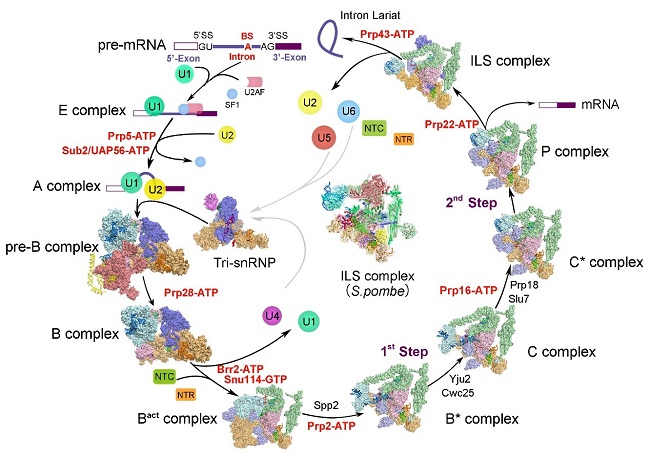
Figure 4. Cryo-EM structure of yeast spliceosome solved by Shi lab.
Prof. Yigong Shi is the corresponding author. Dr. Ruixue Wan (post-doc fellow from the School of Medicine), and Rui Bai (4th year PhD student from the School of Life Sciences) are the co-first authors of this paper. Dr. Chuangye Yan (post-doc fellow from the School of Life Sciences and Center for Life Sciences) and Dr. Jianlin Lei provided technical support on the model building and EM data collection. EM images were acquired at the Tsinghua University Branch of the China National Center for Protein Sciences (Beijing). Data processing was performed on the “Explorer 100” cluster system of Tsinghua National Laboratory for Information Science and Technology, the Computing Platform of China National Center for Protein Sciences (Beijing). This research was funded by the Beijing Innovation Center for Structural Biology and the National Natural Science Foundation of China.
The original link:
https://www.cell.com/cell/fulltext/S0092-8674(19)30155-2
The video link:
https://v.youku.com/v_show/id_XMzc3ODAwNzMxNg==.html
Related publications:
http://science.sciencemag.org/content/early/2016/01/06/science.aad6466
http://science.sciencemag.org/content/early/2015/08/19/science.aac8159
http://science.sciencemag.org/content/early/2015/08/19/science.aac7629
http://science.sciencemag.org/content/early/2016/07/20/science.aag0291
http://science.sciencemag.org/content/early/2016/07/20/science.aag2235
http://science.sciencemag.org/content/early/2016/12/14/science.aak9979.full
http://www.cell.com/cell/fulltext/S0092-8674(17)30954-6
http://www.cell.com/cell/fulltext/S0092-8674(17)31264-3
http://science.sciencemag.org/content/early/2018/05/23/science.aau0325

